Description
Availability
New & Refurbished
Description & Review
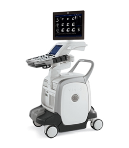
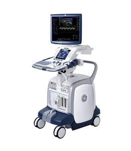
The Vivid S5 is a mid-range, cardiac-focused ultrasound machine that is much smaller, lighter and more reliable than the Vivid 5 it replaced. The Vivid S5 is very similar to the slightly more feature rich Vivid S6 which looks nearly identical to it. Anyone used to the excellent Vivid 7 will find either of these two systems a much more modern upgraded experience with almost all the same features.
Applications
– Cardiac
– Vascular
– OB/GYN
– Abdominal
– MSK
– Urology
– Small Parts
Secondary Applications
– Pediatric
– Breast
– Neonatal
– Transcranial
– Superficial
Features
– 15″ TFT LCD screen
– Coded Harmonics
– ATO [Automatic Tissue Optimization]
– COI [Coded Octave Imaging]
– Confocal Imaging
– Harmonic tissue imaging
– CPI [Coded Phase Inversion]
– DDP [Data Dependent Processing]
– Color Intensity Imaging
– HPRF [High Pulse Repetition Frequency]
– ASO [Automatic Spectrum Optimization]
– TruScan architecture
– CINE Memory
– ECG
– Vascular measurement package
– InSite capability
– iLinq capability
– Image Management and Archiving
– CD-R/DVD-R
– DICOM 3.0
– DICOM Media Support
– EchoPAC Connectivity
– Q-Analysis
– USB Port
– Ethernet port
– 80GB HD
Imaging modes
– B-Mode
– M-Mode
– Color Doppler
– PW Doppler
– CW Doppler
– Duplex
– Dual screen
– Quad screen
– Contrast Imaging
– Tissue Harmonic Imaging
Options
– Anatomical M-Mode
– Smart Depth
– Stress Echo
– OB Application Module
– IMT [Intima Media Thickness]
– Contrast Imaging
– LVO Contrast
– DICOM Network Connectivity
– DICOM Modality Worklist
– DICOM Print
– Database Importation from Vivid 3 and Vivid 4 Systems
– Virtual Printer
– MPEGvue
– Uninterruptible Power Supply


