Description
Description & Review
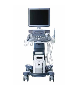
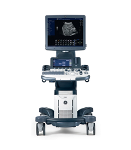
The top of the line Philips iE33 is an excellent shared service ultrasound machine with a high-end set of cardiac and clinical functions. The iE33 combines Philips ease-of-use with the newest possible features and cutting edge imaging.
Applications
– Cardiac.
– Vascular.
– OB/GYN.
– Abdominal.
– MSK.
– Urology.
– Small Parts.
Features
– 17″ or 21″ LCD monitor on articulating arm
– 2 navigation touchscreens
– 3 probe ports
– 57,000 dynamically scalable channels
– Fast system bootup
– Tissue Tracking & Curve Fitting Analysis
– Auto Doppler
– Contrast Imaging
– THI [Tissue Harmonic Imaging]
– TDI [Tissue Doppler Imaging]
– Adaptive Doppler
– Pulse Inversion
– Tissue Synchronization
– Q-Analysis
– xSTREAM image former
– Live Xplane imaging
– SonosCT imaging
– HD Zoom
– Panoramic Imaging
– HPRF [High Pulse Repetition Frequency]
– iSCAN [Intelligent Image Optimization]
– iFOCUS [Intelligent Focusing]
– iOPTIMIZE [TSI, DRS, Flow & Patient optimization]
– iCOMMAND [Voice recognition]
– SonoCT [ Compound imaging ]
– XRES [ Speckle noise reduction ]
– Stress Echo Acquisition Protocol
– Live 3D TEE
– Live xPlane
– ECG/AUX CW
– 2200 frame CINE loop
– QLAB Cardiac 3DQ
– QLAB 2DQ [Cardiac 2D Quantification]
– QLAB IMT [intima-media thickness]
– QLAB SQ [Strain quantification]
– QLAB ROI [Region of Interest]
– TDI [Tissue Doppler Imaging]
– 4 USB ports
– DVD-CD-RW
– 160 GB hard drive
– Netlink DICOM 3.0
– DICOM SR
Imaging Modes
– B-Mode
– M-Mode
– Color Doppler
– Power Doppler
– PW Doppler
– CW Doppler
– 4D
– Duplex
– Triplex
– Dual screen
– Quad screen
– Contrast Imaging
– Tissue Doppler Imaging
– Tissue Harmonic Imaging
Options
– 21″ LCD monitor


