
Description
IBUS 60
Intelligent Breast Volume Ultrasound System
IBUS 60, an intelligent breast volume diagnostic system launched by SIUI, is the third of its kind in the world.
Applied with brand new, cutting-edge ultrasound examination method, IBUS 60 represents a major technological breakthrough in the field of ultrasound imaging and diagnostic mode. Featured with safety and comfort, IBUS 60 offers high-resolution images and reduces missed diagnosis, which is ideal for breast exam especially for one with dense breasts.
Exceptional diagnosis experience
● Nanopure speckle reduction technology
● Intelligent lesion tracking
● Flexible arm control
● Auto scanning
● Comfortable pressure fit
● Efficient workflow
● Unique double coupling
● Mute scanning
● Automatic reset after scanning
● Customized scanning protocol
● Three-dimensional positioning
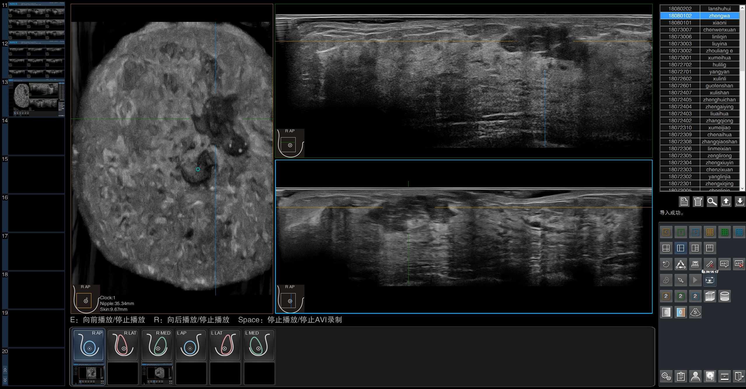
● Breast health reporting system
IBUS VS Traditional Ultrasound
● Standardize the screening, independent from experience and skill
● Enhance detection of tumors, and better reveal the morphology of minute lesions
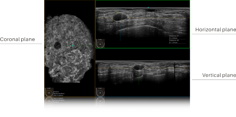
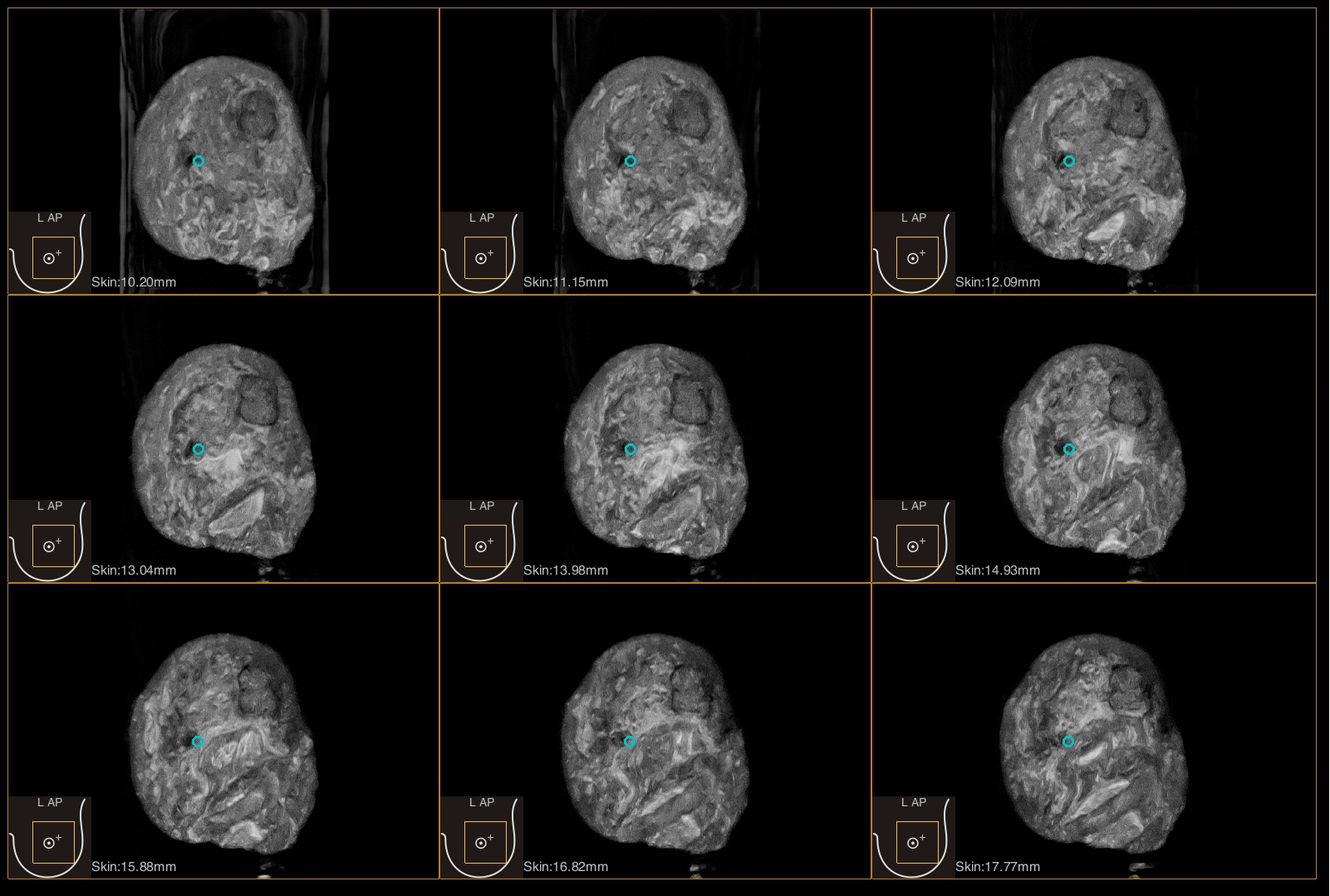
● Free 3D imaging displays transection/coronal plane/sagittal plane, reducing misdiagnosis rate
● Accurate spatial stereo positioning, highly consistent with surgical fields
● Field-of-view is over 3 times larger than traditional 2D probe to capture the large lesions completely
IBUS VS Mammography
● Zero X ray radiation allows rescreening
● More accurate and convenient in diagnosing solid-cystic of tumors
● Higher detection rate of tumors in dense breast
● Display breast with volume ultrasonic tomography
● Easily acquire horizontal, vertical and coronal images
● Accurate spatial stereo positioning, highly consistent with surgical fields
IBUS VS MRI
● Shorten examination time and improve diagnosis efficiency
● Harmless and applicable to all people
● Cost-effective
Full Volume Imaging and Coronal Section with High Clinical Value
Intelligent breast volume ultrasound system acquires data information from multi-section, multi-angle and multi-direction. The coronal scanning intuitively shows the anatomical information of breast tissue in a supine position during operation, which helps surgeon to perform more accurate surgical planning.
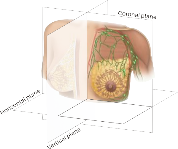
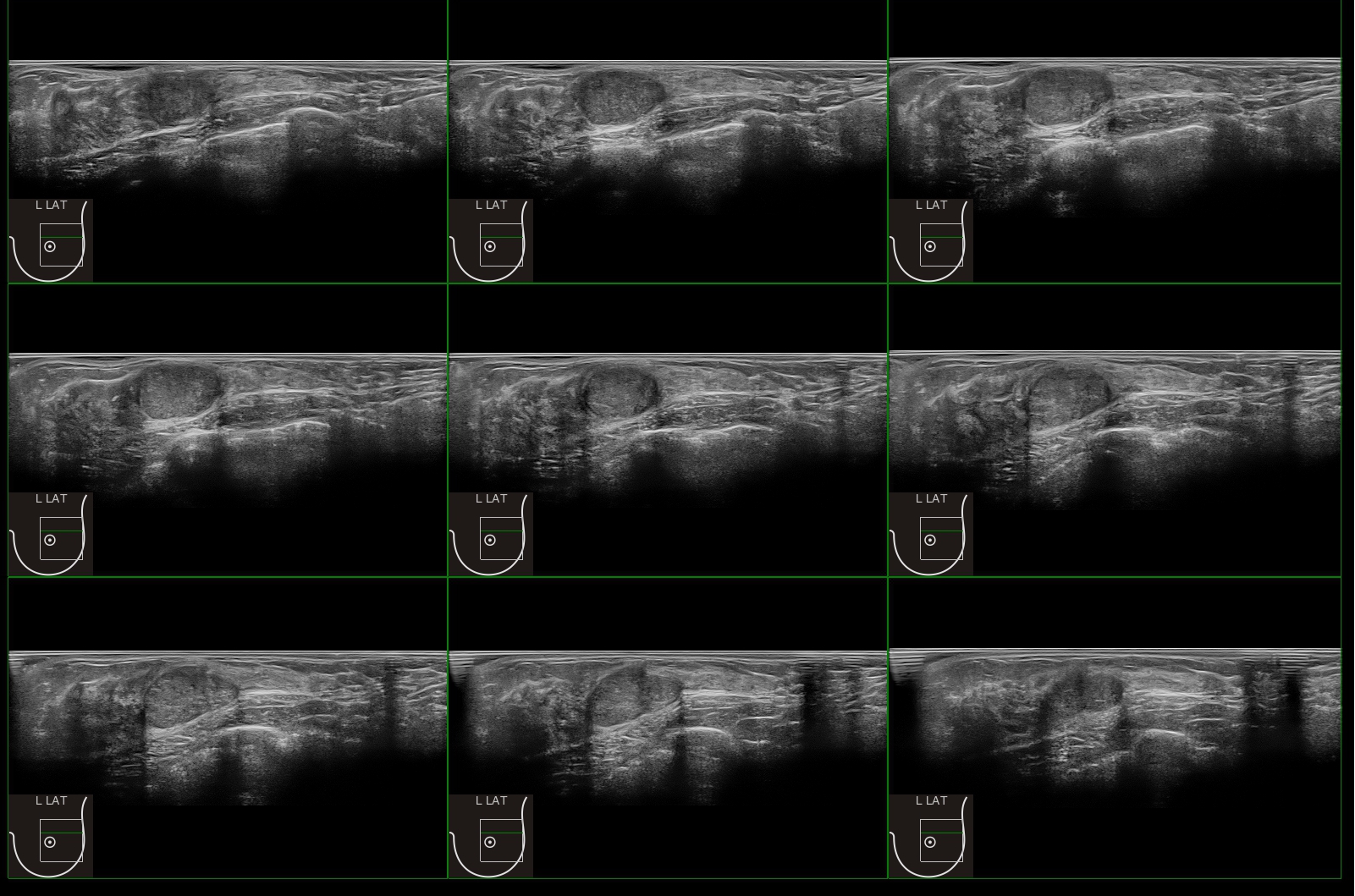
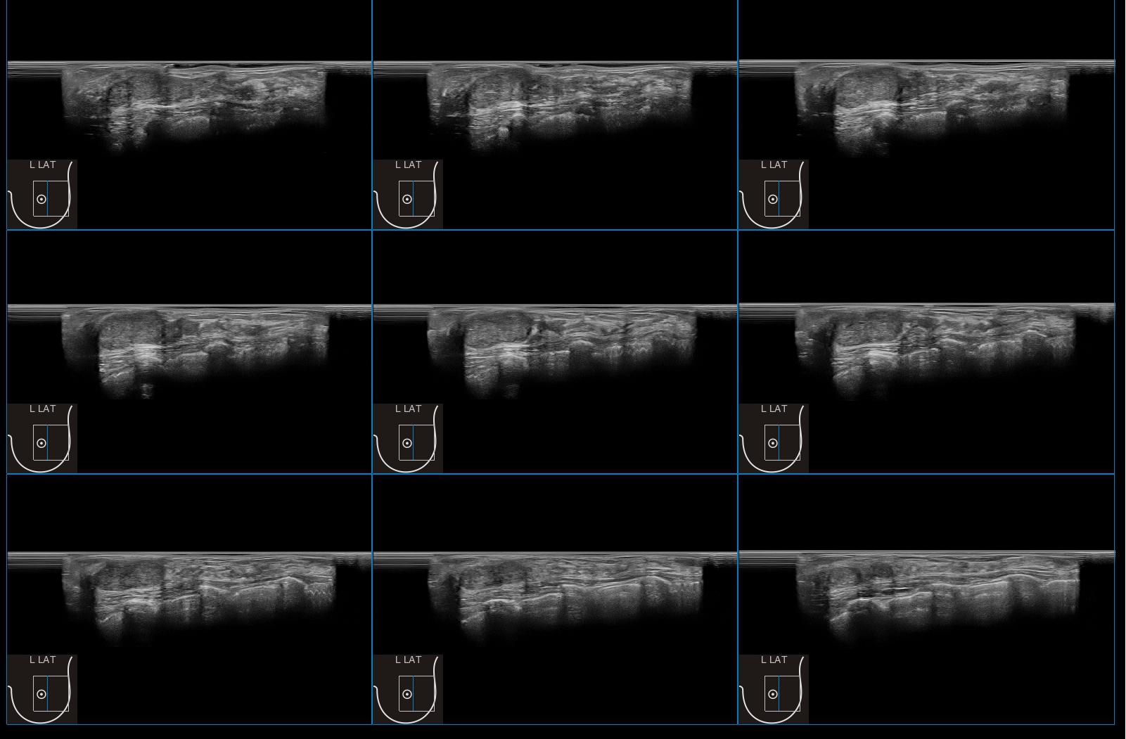
Coronal Plane Vertical Plane


