Description
Description & Review
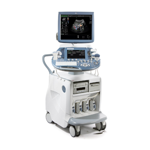
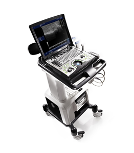
The GE Voluson E8, is General Electric’s top of the line ultrasound machine for the 4D OB/GYN, and women’s health markets. Like all Voluson machines it is a specialist in these categories and offers limited shared service options. Doctors looking for shared service capablities from GE should consider the GE Logiq E9 instead. That said, the E8 is the best in the world at what it does, which is provide incredible, amazing 4D imaging in any probe and situation needed, including the rare 4D linear. The E8 is the successor to the Voluson 730 which has been the most popular and well known ultrasound machines in the world, ever. The E8 improves upon the Voluson 730 in every aspect, screen size, resolution, speed, physical size, and software features. Though very expensive new, refurbished late model Voluson E8’s offer a better value proposition for those not needing HD live, or other features of newer revisions. Voluson E8 BT08-BT10 are good late model revisions. BT12 offers HDlive, and BT13 is the current new revision.
“Top selling system”
Applications
– 3 Cardiac
– 3 Vascular
– 5 OB/GYN
– 5 Abdominal
– 5 MSK
– 5 Urology
– 5 Small Parts
Secondary Applications
– Pediatric
– Neurology
Features
– 15?? LCD monitor (BT06+)
– 10.4? color touchscreen
– 3 Active Probe Ports
– Floating Keyboard: Interactive back-lighting
– BetaView
– Virtual Convex
– Wide Sector
– ATO [Automatic Tissue Optimization]
– Coded Harmonic Imaging with Pulse Inversion Technology
– High Resolution Zoom
– Pan Zoom
– Steering
– Inversion
– Real-time automatic Doppler calcs
– Measurement and Calculations including Worksheets/Report for OB/GYN, Vascular, Cardio, Abdominal, Small-Parts, Urology, Pediatrics, – Musculoskeletal, Neurology
– Multigestational Calculations
– VOCAL II
– HPRF [High Pulse Repetition Frequency]
– SonoVCAD [Matrix array volume technology]
– TruScan [On-board archive]
– VCI [Volume Contrast Imaging]
– SonoAVCfollicle [Sonography-based Automated Volume Count follicle]
– Anatomical M-Mode (BT09+)
– TUI [Tomographic Ultrasound Imaging]
– CE [Coded Excitation]
– XTD Extended View
– SRI II [Speckle reduction imaging]
– CrossXBeam CRI [Compound Resolution Imaging]
– Scan Assistant (BT10+)
– Advanced STIC [Spatio-Temporal Image Correlation] (BT10+)
– STICflow
– SonoRender Start (BT10+)
– SonoNT (BT10+)
– SonoIT (BT13+)
– SonoBiometry (BT13+)
– New Enhanced Rendering with the Dynamic rendering engine (BT10+)
– Elastography (Not available in all Countries) (BT12+)
– Contrast Agent Mode (Not available in all Countries) (BT12+)
– Static 3D Mode
– 4D Biopsy
– Focus and Frequency Composite (FFC) (BT12+)
– Patient Information Database
– Image Archive on hard drive
– 3D/4D data compression
– 512 MB Cine Loop
– Ethernet port
– Video Out
– 6 USB ports
– Integrated HDD (160 GB)
– DVD/CD-RW
Imaging modes
– B-Mode
– M-Mode
– Color Doppler
– Power Doppler
– Spectral Doppler
– PW Doppler
– CW Doppler
– Freehand 3D
– 4D
– Duplex
– Triplex
– Dual screen
– Quad screen
– Contrast Imaging
– Tissue Doppler Imaging
– Tissue Harmonic Imaging
– B-flow
Options
– 19″ LCD monitor (BT08+)
– 4D Real Time
– VOCAL II VCI (Volume Contrast Imaging)
– Advanced VCI and OmniView (BT10+)
– SonoVCAD
– SonoVCADheart [Sonography-based Volume Computer Aided Display] (BT13+)
– SonoVCADlabor
– CW Doppler
– DICOM (BT13+)
– DICOM 4D
– STIC [Spatio-Temporal Image Correlation] (BT09)
– Power Doppler Mode
– CFM Doppler Mode
– HD-Flow Mode
– T.U.I [Tomographic Ultrasound Imaging]
– Coded Contrast Imaging (not available in all countries)
– SonaAVC (BT09+)
– Scan Assistant (BT09)
– Elastography (Not available in all Countries) (BT12+)
– Advanced 4D (BT13+)
– 4D Advanced STIC (BT13+)
– Foot Switch, with programmable functionality
