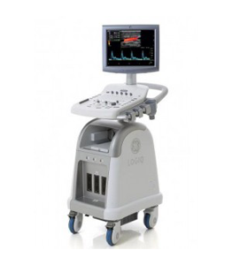
Description
Availability
New
Description & Review
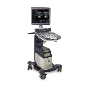
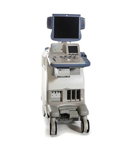
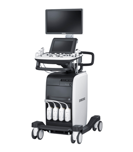
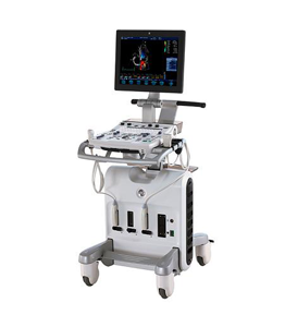
The Voluson S6 offers the right mix of capabilities for enhancing your productivity through extraordinary 2D image quality, volume imaging capabilities, automation tools, easy-to-learn workflow, all combined in an innovative ergonomic design.
– Excellent 4D
– Small & lighter than Voluson 730
“Top selling system”
Applications
– 3 Vascular
– 5 OB/GYN
– 5 Abdominal
– 4 MSK
– 4 Urology
– 4 Small Parts
Secondary Applications
– Pediatric
– Anatomical M-Mode
– HD-Flow
– Color Flow Mode
Features
– 19″ High Resolution TFT LCD Screen
– 3 Active Probe Ports
– High PRF PW Doppler
– Automatic Tissue Optimization
– CE [Coded Excitation]
– CHI [Coded Harmonic Imaging]
– FFC [Focus Frequency Composite]
– CRI [Compound Resolution Imaging]
– CrossXBeam
– Advanced SRI [Speckle Reduction Imaging]
– Virtual Convex
– Wide Sector
– Extended XTD View
– High Resolution Zoom
– Pan Zoom
– SonoRenderStart
– Steering
– Beta-View
– Cine with Dual/Quad display, and Review Loop
– Scan Assistant
– Real-time automatic Doppler calcs
– Measurement Tools and Calculations packages
– Patient information database
– Integrated HD (500 GB)
– 3 USB Ports for External Periphetals
– 2 USB Ports for On-board Peripherals
– Ethernet Port
– HDMI Out Port
– Audio Out Port
Imaging modes
– B-Mode
– M-Mode
– Color Doppler
– Power Doppler
– PW Doppler
– Freehand 3D
– 4D
– Tissue Doppler Imaging
– Tissue Harmonic Imaging
Options
– 3D/4D Advanced
– Advanced VCI with SingleView [Volume Contrast Imaging]
– 4D View PC Software
– DICOM support
– DICOM 3
– XTD
– Anatomical M-mode
– SonoVCADlabor
– Integrated Printers
– DVD Recorder
– ECG Digital Module
– Foot Switch with programmable functionality
